
 Stephanie vL Henkel |
Lipid Biosensors Tackle Toxic Bioagents
|
Lipids are fats and fat-derived substances, distinguished from other organic molecules by their dual oil/water solubility. One part is hydrophobic, the other hydrophilic, and it is this characteristic that allows them to self-assemble. For use as a sensor, the molecules are laid as a double layer ~5 nm thick across the electrodes. But that?s where it gets difficult?lipid bilayers are by nature extremely fragile. So the investigators are evaluating several ways to make them robust enough to survive experiments and to function outside the lab environment.
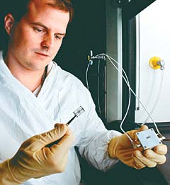
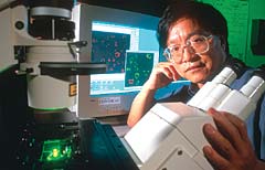
In one configuration, the bilayers are supported on a scaffold built of a thin film of sol-gel and the entire construction is placed on the electrodes. Sol-gel films, formed from a jellylike mass of water, alcohol, and metal oxides, are <1 micron thick but quite durable. Another technique, developed by Sandia?s Darren Branch (photo), uses a hybrid bilayer in which the layer next to the metal electrode is actually not a lipid but an organic silane that attaches to the metal but still supports the upper lipid monolayer.
The researchers are also contending with a second hurdle, that of creating ion ?channels,? or pores formed by proteins in the lipid bilayers that will open and close repeatedly in response to a specific biological agent. In the presence of an agent, the channels change the bilayer?s electrical impedance by permitting the conduction of ions through it. The type and concentration of an agent can thus be identified by measuring the electrical resistance.
When these channels are inserted into lipid bilayers, their ion conduction property closely mimics their activity in living cells. The basic structure of the cell wall is a lipid bilayer, although many other complex structures are also present. Gated ion channel proteins in the cell walls are so sensitive to chemicals that in some cases a single molecule can cause a channel to open or close. The channels will not work unless they are bathed in the lipid bilayer, so they cannot be used as sensors in the rugged polymer matrices used in chemiresistors for VOCs. The solution will ultimately entail attaching antibodies or other molecular recognition molecules to capture the desired molecules on the ion channels, and this could turn out to be the thorniest problem of all.
Contact Darren Branch, Sandia National Laboratories, Albuquerque, NM; 505-284-5843, [email protected].
Coated GaAs Wafer Detects Neutrons
 A very tiny detector under development at Argonne in a project headed by Raymond Klann is built around a gallium arsenide wafer which, when coated with boron or lithium, can detect neutrons such as those emitted by the fissile materials that fuel nuclear weapons. The device could help international inspectors keep concealed nuclear weapons and materials out of this and other countries, and it could also find work in high-vacuum space applications.
The wafers require <50 V, operate at room temperature, can withstand relatively high radiation fields, and do not degrade over time. The coating is key?neutrons striking it produce an easily detectable cascade of charged particles. The wafers, made with conventional microchip processing techniques by coinventor Doug McGregor of Kansas State University, can be tailored for specific applications by varying the type and thickness of the coating. The researchers also discovered that etching the wafer with cylindrical holes increased the detector?s sensitivity, and are experimenting with various coatings and combinations of hole sizes and spacings.
The work was funded by the U.S. DoE?s Office of Science.
Contact Raymond Klann, Argonne National Laboratory, Argonne, IL; 630-252-4305, [email protected].
Doped Polymer Discs Sniff Decay and Change Color
The Fearmonger News Service has got most of us worked up about what we eat?the wrong stuff and too much of it. But the general assumption is that by judicious selection and moderate intake we can elude obesity and other maladies attributable to a diet heavy on the fat, sugar, and salt. If only it were that simple.
While we know better than to eat something green and furry from the back of the fridge, not everything goes bad from the outside in. A sniff test might advise against the frozen shrimp we just brought home, but the olfactory sense can be put out of commission by a head cold or advancing years. And besides, who can smell anything that?s both frozen and sealed under layers of plastic wrap?
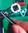
Dwight Miller and his colleagues at the National Center for Toxicological Research have come up with an elegant way to positively identify even the early onset of food spoilage?polymer discs doped with complex organic dyes. Intended for inclusion in packaged foods  sold in the market, these discs change color in the presence of vapors characteristic of decay. The color is determined by the type of food being monitored, and reference colors are printed on the package.
Take those shrimp, for instance. The disc goes from a safe yellow to a warning blue in the presence of trimethylamine, dimethylamine, and ammonia exuded by rotting fish. (In the photo, the yellow disc is on the outside of the package and the blue is inside.) The same holds true of beef and lamb (and elk, according to Miller). Next on the researchers? agenda are frozen vegetables, whose formic, acetic, and other acid emissions of decay will be assigned their own advisory hues.
Contact Dwight Miller, National Center for Toxicological Research, Jefferson, AR; 870-543-7319, [email protected].
Ammunition Tags Trace Firearm History
 When a firearm is discharged, the ammunition picks up assorted scratches and dings that can help criminal investigators match the bullet to the weapon from which it was fired. This is all well and good?if you have the candidate weapon and can do a test firing. But most people who shoot with criminal intent don?t leave their guns behind, and the telltale marks can change with time or type of ammunition.
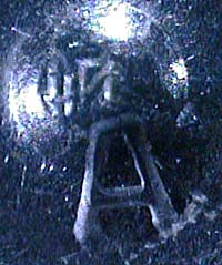
NanoVia has devised a sure-fire way not only to connect a gun with a recovered bullet, but also to trace the weapon?s ownership history. With the NanoTag technique, an embossing surface integrated into the inside of a firearm stamps each cartridge casing as bullets are fired. The resulting mark is a unique, indelible, microscopic code in the form of encrypted symbols, bar codes, or simple 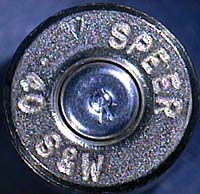 alphanumeric codes (such as the make, model, and some type of tracking number) that can be accessed at the manufacturer?s level to reveal the serial number and purchase history of the gun.
Because the code can be read by imaging equipment available in most forensic labs, no test firing is necessary. Moreover, NanoTag ballistic identification does not require national registration of new firearms or cross-jurisdictional databases of ballistic ?fingerprint? images of the weapons.
A number of state-level justice departments and federal agencies are interested in the NanoTag technology. The California Department of Justice has begun an evaluation program designed to optimize and test various code configurations and placement sites for the embossing surface in assorted types of firearm for forensic value and repeatability, and to establish guidelines for implementation.
Contact Todd Lizotte, NanoVia, LP, Londonderry, NH; 603-421-0713, [email protected].
Surface Plasmon Resonance Speeds Chemical Analysis in the Field
 Chemical detection in the field could get easier with a novel surface plasmon resonance (SPR) probe developed at the Westinghouse Savannah River Co. SPR is an optical phenomenon that enables the detection of molecules adhering to a surface.
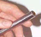
The researchers took a commercially available fiber-optic attenuated total reflection (ATR) probe and modified it by depositing a series of thin layers of gold and silica on the probe?s hemispherical lens. Molecules adsorbed onto the silica are detected by a loss of reflected light at a specific frequency or wavelength as the incident light is coupled into the surface plasmon wave.
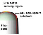 Using an ATR probe as the starting optics allows the sensor to be placed at the end of a fiber-optic cable that carries both the incident light and the reflected signal back to the analyzer. The curvature of the lens compensates for the divergence of the source fiber and refocuses the light back to the collection fiber.
The sensor has been shown to produce high-resolution, low-noise SPR spectra on a par with benchtop equipment. Extremely sensitive to water vapor adsorption, it has demonstrated sub-ppm sensitivity over a range of ~0%?100% RH (0 to >20,000 ppm at 25°C, 1 atm.) and can be used as well to measure any gas-phase species that can condense on the probe tip. The probe would be particularly helpful in applications where an electrical measurement is undesirable, or where there is EMI.
Contact Eric Frickey, Westinghouse Savannah River Co., Aiken, SC; 803-725-0406, [email protected].
Improved Molecular Beacons Illuminate Cancer, Viruses
 A new class of fluorescing molecular beacons under development at Georgia Tech and Emory could facilitate the detection of cancer and viral infections both in vivo and in the lab. The beacons consist of a fluorescent dye molecule and a quencher molecule, whose function is to prevent fluorescent emission from the dye, on opposite ends of an oligonucleotide engineered to match specific genetic sequences associated with disease.
Dye and quencher are initially held close together in a hairpin shape. Delivered into cells, the beacons seek out matching sequences in genetic material known as messenger RNA (mRNA). If the beacons encounter and bind with their targets, Watson-Crick base pairs holding the dye and quencher together break and allow the emission of a specific fluorescent signal when excited by light. Research team leader Gang Bao and colleagues Andrew Tsourkas and Phil Santangelo believe the technique to be the first for imaging RNA in living cells.
There is a problem, though. A cell?s normal enzyme activity can separate the dye from the quencher and produce a false positive signal. The researchers? solution is a system in which two beacons attach to the same target mRNA on adjacent binding sites. When that happens, fluorescence resonance energy transfer (FRET) between a donor beacon and an acceptor beacon creates a red-shifted optical signal that can be distinguished from false signals produced by the enzymatic digestion of single beacons. Because FRET is extremely sensitive to the distance between donor and acceptor molecules, the transfer occurs only when the donor and acceptor molecules are bound to the same mRNA target.
The investigators are also working on beacons containing magnetic nanoparticles for use in body tissues too deep for optical imaging to work. When two beacons attach to adjacent sites of a target mRNA, the disturbance they create in water molecules can be detected with magnetic resonance imaging. Accordingly, the researchers have combined the nanoparticles with oligonucleotides necessary for recognizing and binding to target mRNA. The ideal diagnostic system might eventually consist of optical beacons for cellular studies outside the body, a delivery scheme and additional technologies for shallow tissue imaging, and magnetic beacons for deep-tissue imaging.
Because a definitive cancer diagnosis requires the recognition of several markers, future generations of the detection system will probably incorporate multiple beacons, each targeting a different marker and producing a unique signal. Because the beacons specifically attach to mRNA, they could perhaps slow down or halt the growth of cancer cells. It will, of course, have to be shown that they don?t harm healthy cells.
Also being investigated are a way to target specific organ systems, more rapidly disperse the beacons into cells, and recognize genetic sequences that signify the presence of viruses and allow their detection in hours rather than days. The basic technology could also prove useful for drug discovery and parmacogenomics.
The research is supported by the Wallace H. Coulter Foundation and the National Science Foundation.
Contact Gang Bao, Wallace H. Coulter Dept. of Biomedical Engineering, a joint operation of Georgia Tech and Emory, Atlanta, GA; 404-385-0373, [email protected].
Now We Know
 For the following to make any sense, please see the R&D department in the February 2003 issue of Sensors (?Gait Recognition Enters Biometrics Field?). A faithful reader has enlightened us as follows:
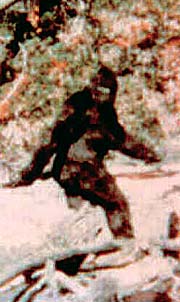
The Sasquatch in the film has always been known to be female because of rather obvious breasts. Her ?real identity? is now known because a family has come forward and identified their now-deceased father as the practical joker and his still-living wife as the hairy woman in the pictures?she and several kids were shown on TV answering questions with a clip of the film.
Apparently he started with fake feet, which [the family] showed, and then did the movie. It went a lot further than he expected, but whenever evidence waned, he went out and did some more footprints and then let other people find them. He stayed pretty much away from the spotlight.
Mike Firth, [email protected]