
The capacitively coupled noncontact electrode (CCNE), a new device that measures electrocardiogram (ECG) interbeat intervals through clothing, underwent a 40-person human trial at the Walter Reed Army Institute of Research (WRAIR) this past year. This sensor can detect ECG and respiratory signals through clothing and is being considered by the U.S. Army as a physiological monitoring detection sensor on the Future Force Warrior System uniform and for en route care to monitor wounded soldiers during evacuation. In this article, we compare three CCNE sensors to an FDA-approved monitor (using contact electrodes) to determine if the R-R interbeat intervals (the time between two consecutive R waves of the cardiac cycle and used to determine heart rate) measured by the two methods are the same or not. We present the results from determining heart rate based on interbeat intervals of the ECG while the test subject is at rest in a supine position.
 |
Background and Rationale
Various attempts have been made to overcome the limitations of wet electrode technology for measuring bioelectric signals such as ECG and electroencephalogram (EEG) on the human body. Advances include a class of surface electrodes that doesn't use electrolytes [1]; these are referred to as active electrodes and use an impedance transformation at the sensing site via active electronics. Active electrodes are subdivided into two types: dry electrodes, which rely on a metallic surface in direct contact with the test subject and use a combination of resistive and capacitive coupling to the local skin potential; and insulated electrodes, which use only capacitive coupling [2].
In 2001, researchers at Quantum Applied Science and Research (QUASAR) developed a new class of sensor that measures the electric potential in free space, i.e., without physical contact to any object. These sensors were able to measure the ECG of a fully clothed person standing within a range of about 25 cm. In 2002, QUASAR developed a compact version of the sensor and named it the capacitively coupled noncontact electrode (CCNE), specifically to measure ECG through clothing (see Figure 1). This sensor could also be mounted in a litter configuration, eliminating the need for contact electrodes and providing early monitoring of wounded soldiers during evacuation.
For the first human trial of this new electrode technology, our goal was to compare the CCNE operating through regular clothing with a conventional 3-lead ECG using resistive electrodes to measure interbeat intervals.
The principal application envisioned for this technology is continuous monitoring of the ECG of military personnel as part of the Warfighter Physiological Status Monitor (WPSM) to be contained within the Future Force Warrior (FFW) soldier ensemble. FFW is the Army's flagship science and technology initiative to develop and demonstrate revolutionary capabilities for Future Force soldier systems. The many problems associated with contact electrodes for long-term ECG monitoring include loss of contact to the test subject due to drying of application glue or to environmental factors (e.g., rain) as well as test subject resistance to wearing the electrodes due to discomfort (e.g., skin irritation). Therefore, a noncontact ECG system would be of great benefit to the Army. Integrated into the FFW uniform, a noncontact system would provide not only simple heart-rate changes and interbeat interval variability, but with sufficient waveform fidelity, could also monitor arrhythmias and conduction abnormalities associated with hypotension and cardiac ischemia. A combat medic armed with the information from such a system could properly ascertain the level of injury and the survival potential of the injured and provide optimum care in the battlefield and during en route care from the battlefield.
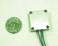 Figure 1. This photo shows the capacitively coupled noncontact electrode (CCNE) used in this study. This first version of the ECG electrode, including all amplification electronics, is ~1 in. square and 0.35 in. thick. |
Methods and Materials
The Walter Reed Army Institute of Research Clinical Trials Division recruited test subjects for five weight groups of four test subjects each; one set of five groups was female and another set male, for a total of 40 test subjects. To be included in the study, volunteers were required to be healthy males and females with no known heart defects, who ranged in weight from 101–173 lb. for females, and 124–220 lb. for males. These weight ranges represent the ninety-fifth percentile of the men and women in the Armed Forces today. A pilot study with two test subjects determined that, for this initial trial, the best results were obtained by placing sensors 4 in. below the right and left nipples, and one on the left shoulder blade, dorsal to the other two sensors [3]. Because the data acquisition system and test subject were separated by about 2 m, the test subject wore a ground strap attached to the right wrist to minimize DC potential differences. This ground strap has been eliminated in the latest iteration of the technology. A commercially available Velcro strap (see Figure 2) was used to hold the sensors to the body over a cotton T-shirt provided for the study (see Figure 3).
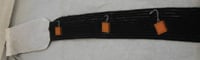 Figure 2. A commercially available Velcro strap was used to hold the sensors. Here we see the arrangement of the sensors on the strap. |
Each sensor detects the local electric potential measured relative to another voltage. For conventional electrodes, the reference voltage is usually obtained by a resistive connection to one or more limbs. The goal of the CCNE approach is to operate through clothing without a resistive electrical contact to the test subject. Accordingly, the output of each CCNE electrode was recorded relative to one of the other CCNE electrodes, giving a total of three purely capacitive difference signals, i.e., channel 1 – channel 2 (X0-X1), channel 1 – channel 3 (X0-X2), and channel 2 – channel 3 (X1-X2).
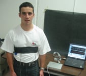 Figure 3. Here we see a QUASAR employee wearing the T-shirt and Velcro strap. |
Each test subject was connected to a commercial clinical ECG monitor, pulse oximeter, and blood-pressure cuff. An FDA-approved clinical ECG monitor (the BCI Model #3404-001 AutoCorr Plus) was used to provide the "gold standard" reference ECG waveforms for all comparisons with the CCNE waveforms. The frontal plane configuration for the 3-lead ECG was used to take resistive measurements [4] with the three contact electrodes placed on the right/left chest and lower abdomen. The test equipment was connected to a data acquisition system by using a Measurement Computing PC card connected to a laptop computer running LabVIEW. Data were sampled at 500 Hz and stored digitally on the laptop, which was running on battery power. The test subject was put into the supine position and data were taken for 1 min. with normal breathing.
Study Results
We applied a peak finding algorithm to detect the R wave maximum of the ECG in each of the waveforms and the time between two consecutive peaks was calculated in milliseconds. The resulting interbeat interval was used to compare the CCNE results with those from the conventional ECG contact electrodes. The raw waveforms for one test subject as seen in Figure 4 show that the two waveforms are very similar in nature for this individual. This result is typical of most of the test subjects.
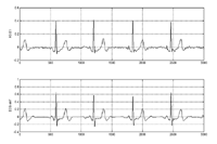 Figure 4. This graph compares CCNE difference signals with those generated using contact ECG electrodes. |
The interbeat intervals were calculated and compared. A typical result for one test subject using this analysis is shown in Figure 5. This shows that the interbeat intervals from the CCNE and the resistive-contact electrodes look identical.
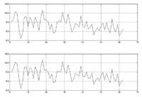 Figure 5. This graph compares the R-R interbeat intervals of CCNE and ECG electrodes on data taken for 1 min. |
For each test subject, we performed Pearson's correlation between the CCNE difference signals and the interbeat intervals obtained from the ECG contact electrodes, with the results shown in Figure 6. We can see from these results the high degree of correlation between the three difference signals of pairs of CCNE electrodes and the ECG contact electrodes.
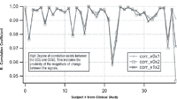 Figure 6. A graph of the correlation between difference signals of CCNE with ECG contact electrodes shows a high degree of correlation between the CCNE and ECG contact-electrode interbeat intervals. |
The high degree of correlation suggests the two sets of intervals are nearly identical; however, a high degree of correlation does not exclude the possibility that the CCNE intervals are biased (i.e., the CCNE intervals might be consistently higher or lower than the corresponding ECG intervals). To determine whether the CCNE intervals were significantly biased compared to the "gold standard" ECG intervals, we looked at two properties of the CCNE intervals: "bias" and "closeness" (distance of CCNE intervals from the ECG intervals). Examining the difference scores (i.e., the difference signal measurement minus the corresponding ECG measurement), we found that two of the three CCNE difference signals, X0-X2 and X1-X2, gave unbiased estimates for all 39 test subjects, and the CCNE difference signal X0-X1 gave unbiased estimates for 38 out of 39 test subjects (test subject 8 was removed from the study due to data acquisition problems with that test subject). The only biased estimate came from subjects 24 and 34 (females in weight class 5).
 Figure 7. This photo shows the latest version of the improved CCNE. |
To see how close the CCNE difference signal interbeat intervals were to the ECG contact electrodes' interbeat intervals, we looked at the absolute value of the differences between the difference signal Xi-Xj interbeat interval (in milliseconds) and the corresponding ECG contact electrodes' interbeat interval (in milliseconds). For 20 out of 39 test subjects all CCNE difference signal interbeat intervals were within 10 ms of the corresponding ECG contact electrodes' interbeat interval. For 30 out of 39 test subjects all CCNE difference signal interbeat intervals were within 14 ms of the ECG contact electrodes' interval. Three of the nine test subjects with absolute differences exceeding 14 ms were females in weight class 5.
To determine if the three CCNE difference signals gave similar results for each test subject, we checked the proportion of positive, negative, and zero difference scores for each test subject. When we applied a chi-square test with four degrees of freedom for homogeneous proportions for (3-by-3) contingency tables, we found that 37 out of 39 test subjects had difference signals giving statistically identical results. The only two test subjects (24 and 34) having dissimilar results were both females in weight class 5.
Other results shown were: 1. Within each weight class, females showed more variability than males; 2. Males and females in weight class 5 showed the largest relative measures of variability (i.e., had the largest chi-square values); and 3. Within each gender, weight class 1 showed the smallest relative measures of variability.
Discussion and Conclusions
Although resistive contact electrodes for measurement of ECG are the gold standard [5], new methods are becoming available to accomplish this task. In this study, capacitively coupled (through clothing) sensors were evaluated against this standard to determine if the R-R interbeat intervals of the two methods were the same or not. The test results indicate that, relative to the ECG contact electrode, the CCNE sensors work reliably for determining R-R interbeat intervals. Statistical analysis of the test results between the CCNE and the contact electrode give the following conclusions:
1 The CCNE method gives "unbiased" estimates (116 out of 117 difference signals gave "unbiased" estimates).
2 The CCNE method gives estimates close to the ECG measurements (30 out of 39 test subjects had all CCNE intervals within 14 ms of the ECG intervals).
3 The CCNE difference signals give statistically similar results within each test subject (37 out of 39 test subjects had statistically similar results).
4 Females showed more variability than males for each weight class.
5 Males and females in weight class 5 had the largest measures of variability.
This study has shown that we can reliably obtain ECG interbeat intervals through clothing using the new noncontact capacitively coupled electrodes. More work is required to study the fidelity of the waveforms to each other to determine if the waveform fidelity is sufficient for interpretive ECG analysis for clinical diagnosis. This work produced valuable information which led to the development of a more robust sensor, shown in Figure 7. Since this study was done, a wireless solution has been developed and tested. Refinements to the technology have resulted in high-fidelity ECG waveforms. Further work is currently underway.
Material has been reviewed by the Walter Reed Army Institute of Research. There is no objection to its presentation and/or publication. The opinions or assertions contained herein are the private views of the authors, and are not to be construed as official, or as reflecting true views of the Department of the Army or USUHS or the Department of Defense.
This information was presented at the NATO Research and Technology Organisation (Human Factors & Medicine Panel) meeting in cooperation with the Advanced Technology Applications for Combat Casualty Care conference in St. Petersburg, FL, August 16–18, 2004. Special thanks to the Defense Advanced Research Projects Agency (DARPA).
Jaime M. Lee, M.S.E. (301-619-4501, [email protected]), Frederick Pearce, Ph.D. (301-319-9761, [email protected]), and Craig Morrissette, Ph.D. (301-319-9969), can be reached at U.S. Army Medical Research and Materiel Command (MRMC); Andrew D. Hibbs, Ph.D. and Robert Matthews, Ph.D., can be reached at Quantum Applied Science and Research (QUASAR), San Diego, CA; 858-373-0231, [email protected], [email protected].
References:
1. Richardson, P.C., "The insulated electrode: A pasteless electrocardiographic technique," Proc. Annu. Conf. Eng. Med. Biol., 1968, 9:15.7.
2. Prance, R.J., "An ultra-low-noise electrical-potential probe for human-body scanning," Meas. Sci. Technol. Vol. 11, 2000, pp. 1–7.
3. Rawlings, Charles A., Electrocardiography, 1993, pp. 30–31.
4. Ibid., pp. 27–28.
5. ANSI/AAMI Standard – EC12:2000, revision to EC12:1991 Disposable Electrodes, pp. 1–14.